Time Resolved Cathodoluminescence (TRCL)
Cathodoluminescence (CL) is a powerful spectroscopic technique: it consists in the analysis of the light emitted by a material excited by a focused electron beam. It can be realized in both Scanning and Transmission Electron Microscopes (SEM and TEM respectively), taking advantage of nm (sub-nm) spatial resolution: its complementarity with standard micro-photoluminescensce makes CL a fundamental tool in many domains such as mineralogy/planetary science[1], material science[2], plasmonics[3], nano-photonics [4,5] and even biology [6].
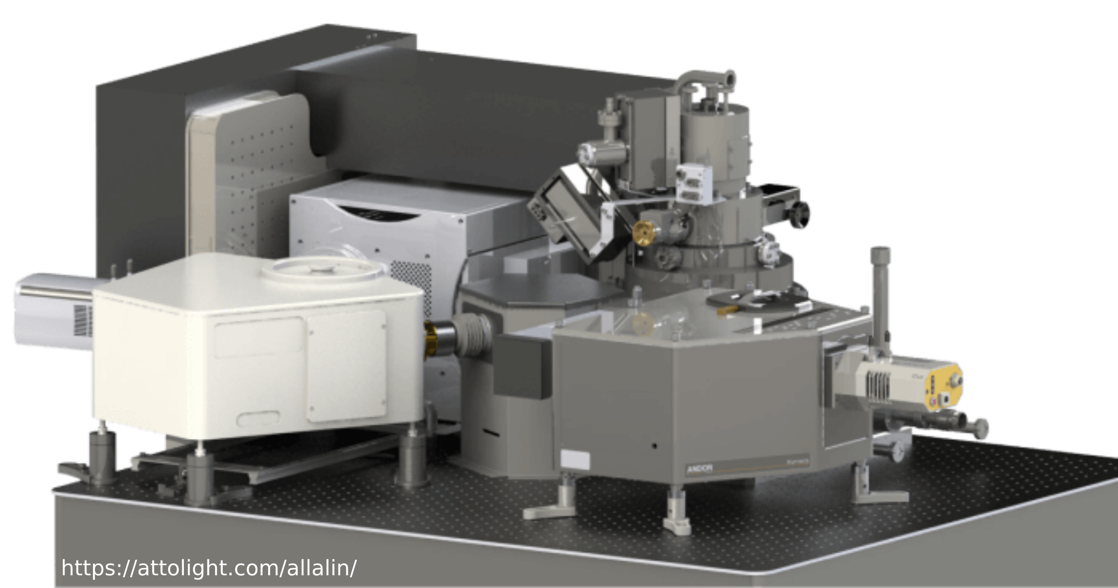
CL spectroscopy was born in standard SEM microscopes, by adding a piezo-controlled parabolic/ellipsoidal mirror[7] to collect the luminescence from the excited sample. Many group around the world still use this approach [2], with a variety of movable collection optics from microscope objectives (both refractive and reflective) to optical fibers allowing numerical apertures as high as 0.95 and a great measurement flexibility. Our commercial tool on the contrary include a fixed reflective mirror to collect the light with a reasonable high numerical aperture (0.72 in air): this feature allows quantitative cathodoluminescence, i.e. the comparison of measurements from different sessions. The tool, Chronos, has been developed by the swiss company Attolight [8]: it allows classic scanning SEM CL spectroscopy as well as time-resolved CL measurements, thanks to a picosecond laser triggering the electron emission from the gun.
Chronos has been installed at C2N in November 2015, and upgraded in 2019 by the SUNLIT team (S.Collin) , and it became part of the PANAM platform in 2021.
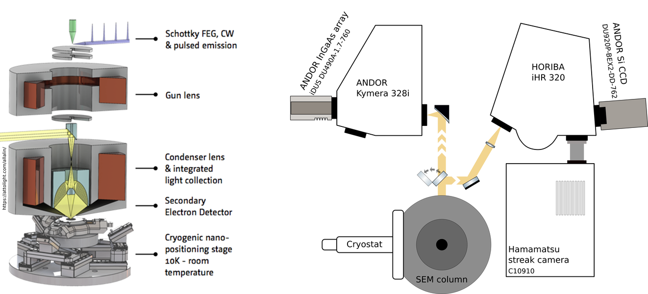
Allalin/Chronos injection and collection paths. From https://attolight.com/allalin/
References
[1] Cathodoluminescence and its Application in the Planetary Sciences, editor Gucsik A., Springer Berlin Heidelberg, 2009, doi:10.1007/978-3-540-87529-1.
[2] T. Coenen and N. M. Haegel, Cathodoluminescence for the 21st Century: Learning More from Light, Applied Physics Reviews 4, 031103 (2017).
[3] A. Losquin and T. T. A. Lummen, Electron Microscopy Methods for Space-, Energy-, and Time-Resolved Plasmonics, Front. Phys. 12, 127301 (2017).
[4] C. Roques-Carmes et al., Free-Electron–Light Interactions in Nanophotonics, Applied Physics Reviews 10, 011303 (2023).
[5] Z. Liu et al., Scanning Cathodoluminescence Microscopy: Applications in Semiconductor and Metallic Nanostructures, Opto-Electronic Advances 1, 18000701 (2018).
[6] Y. Nawa et al., "Dynamic and high-resolution live cell imaging by direct electron beam excitation," Opt. Express 20, 5629-5635 (2012).
[7] J. B. Steyn, P. Giles†, and D. B. Holt, An Efficient Spectroscopic Detection System for Cathodoluminescence Mode Scanning Electron Microscopy (SEM), Journal of Microscopy 107, 107 (1976).
[8] https://attolight.com/
Attolight Allalin/Chronos Time Tesolved CathodoLuminescence
Continous CL
Time-Resolved CL
Secondary Electron Microscopy (SEM)
Electron Beam Induced Current (EBIC)
electron energy range: 3 keV -10 keV
e-beam pulse width: 10 ps
achromatic collection optics: reflective objective
spectral range: 250 nm - 1.7 µm
sample temperature: 5 K - 300 K
streak camera
APD, PMT and TCSPC
ultra stable sample stage (drift < 300nm/h)